


Symptoms on Chilli
- Organs attacked :
| Leaves | Rod |
- Symptoms :
- Initially small circular to elongated chlorotic lesions (Figure 1).
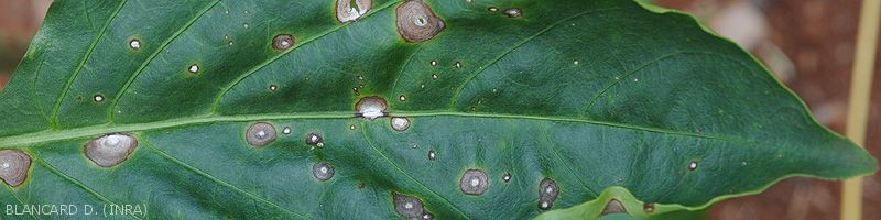
- Subsequently, the spots expand, turn brown, and gradually become necrotic (figure 2). A reddish-brown border encircles them. their diameter can be more than 1 cm.
- The center of the spots lightens gradually taking on a gray to whitish tint, and sometimes presents concentric patterns giving them a so-called "frog eye" appearance. A yellow halo is sometimes visible (Figure 3).
- Similar but elongated lesions are sometimes visible on the petioles (Figure 4) and the stem.
- Degraded fabrics dry out, split and eventually fall off; the leaf blade is thus partially riddled.
- Severely affected leaves turn yellow, wither, and may drop.
- Signs : Fruiting bodies of the fungus dot the lesions, sometimes giving them a slightly light gray tint. Under the light microscope, many conidiophores can be observed carrying hyaline, elongated and septate conidia.
- Possible confusion : rare
- >>> More pictures
- Affected production areas :
| Mayotte | New Caledonia |
- Cercospora spp. reported on pepper : Cercospora capsici Heald & FA Wolf (syn. Cercospora unamunoi, etc.), Passalora capsicicola (Vassiljevsky) U. Braun & FO Freire (syn. Cercospora capsicicola , etc.).
Figure 1
Figure 2
Figure 3
Figure 4